TEMNOGRAPHY

Applications

How the Temnograph Encephalic Subcranial (TES®) works, and the clinical situations where it is used.

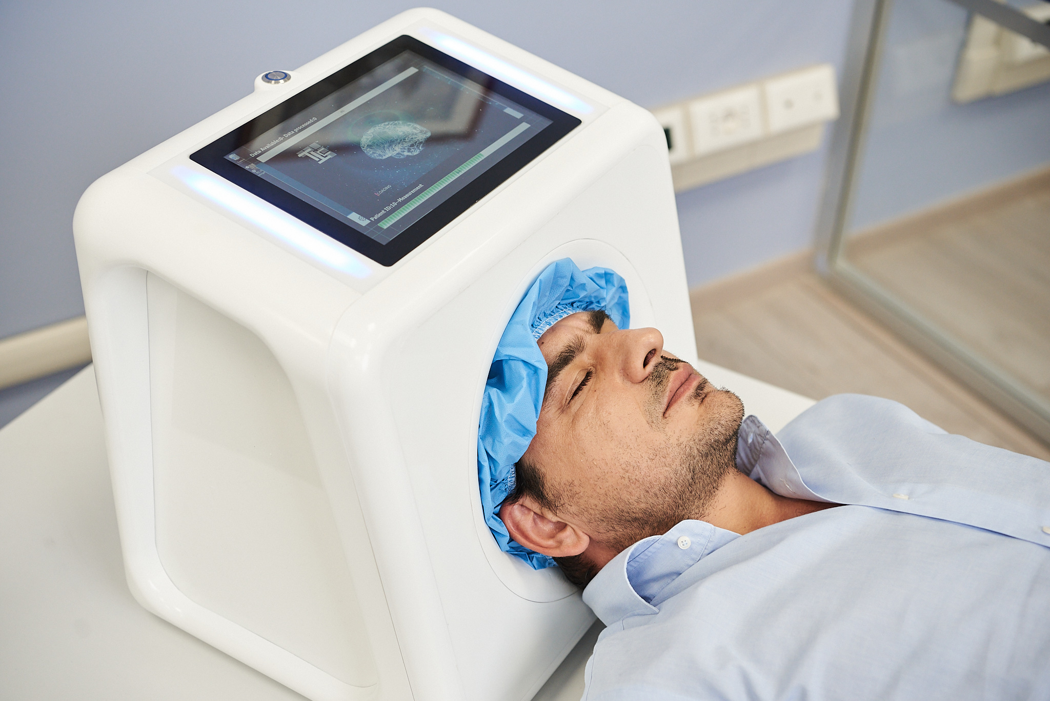
A patient undergoing a TES scan, with live scan data shown on the device screen

The technology

How TES® works

Tissue and detection

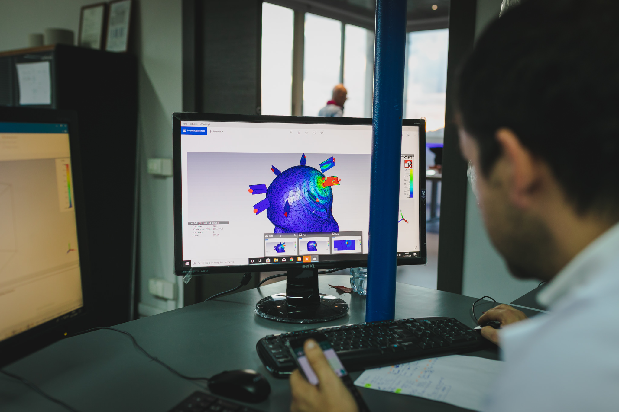
The electromagnetic fields, generated by microwaves, propagate through tissues undergoing complex alterations generated by reflections, refractions, and phase angle changes, considered in both the time and space domains. These perturbations alter the characteristic imprint of the healthy brain and are captured by the Temnograph, which uses them as a radar to determine the presence or absence of the sought-after abnormality.

Safe

TES is non invasive, does not employ ionizing radiation and the exam is repeatable when clinically appropriate. TES utilizes very low-power RF energy; the intensity and duration of exposure are kept at minimal levels to support the safety of both patients and operators.

Fast and Easy

TES technology, through its detection process, directly yields the exam result as a simple “YES” if the target abnormality is present or “NO” if it’s absent. The result does not require interpretation by an expert user to read, and shows the estimated location where detected.

A century of diagnostic imaging

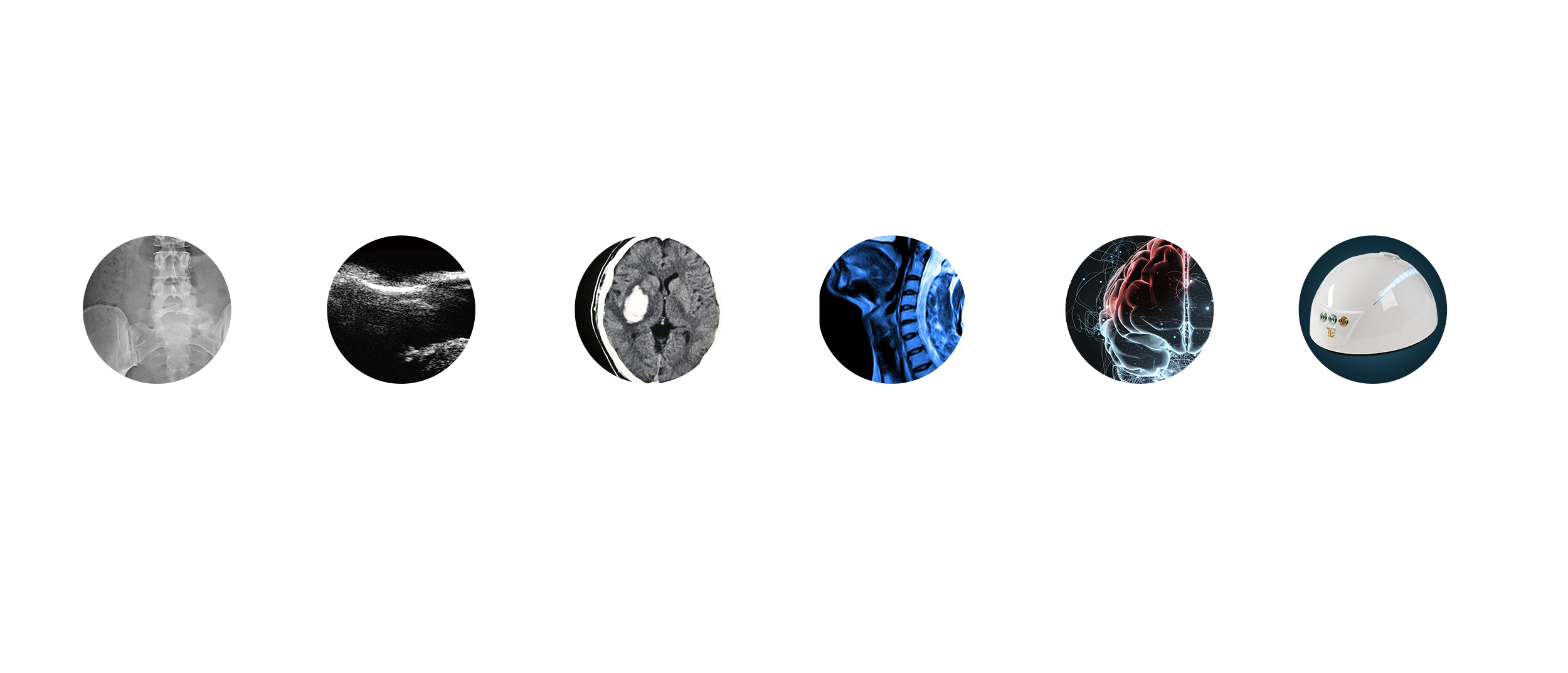
Timeline of diagnostic imaging: 1895 X-ray, 1950 echography, 1967 CT scan, 1978 MRI, 2016 Temnography®, 2018 the first Temnograph Encephalic Subcranial (TES)

Where it is used

Clinical applications

Wherever a fast, repeatable answer on intracranial bleeding changes what happens next.

Stroke
01Current Clinical Focus

Stroke: a fast bedside assessment to help identify haemorrhage.

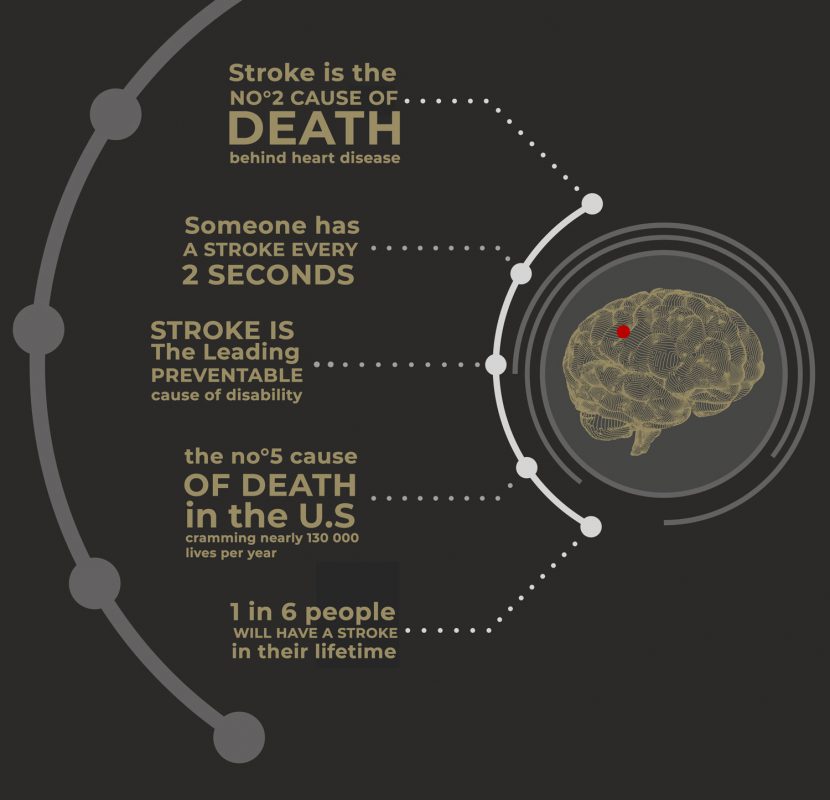
There are two types of stroke: haemorrhagic and ischemic, with two different paths of therapy. When a stroke happens, quick treatment is crucial.

A lot of time is wasted waiting to arrive at hospital and make CT or MRI, in order to decide where to transfer the patient or when to start therapy.

So the first step on the care pathway is diagnostic assessment.

But in case of stroke each minute is crucial.

With TES® it's possible to go directly to the patient's location and complete an approximately 5-minute standardized bedside assessment: whether a haemorrhage is present or not — so emergency (pre-hospital) teams know immediately where to bring the patient and can start therapy without delay.
TES device used to support stroke triage

Without oxygen-rich blood, brain cells die.

Temnography TES® — supporting faster triage when every minute counts.
Thrombolytic Therapy
02Clinical Development

Thrombolytic Therapy (tPA): a potential monitoring application for unwanted bleeding as a side effect.

The protocol during thrombolytic therapy provides for the execution of a control CT scan to determine if there is the occurrence of cerebral bleeding as a side effect.

This examination is usually done after 24 hours.

In clinical development, TES® is being explored as a way to monitor for bleeding at any point after thrombolytic therapy begins, without the delay of scheduling a follow-up CT or adding further ionizing radiation.

TES® uses non-ionizing, very low-power RF energy and is repeatable when clinically appropriate.

Head trauma
03Current Clinical Focus

Head trauma, identifying whether there is a brain hemorrhage or not.

In case of trauma, it is necessary to know in the shortest possible time if the patient is having a cerebral hemorrhage, in order to establish where to transport the patient and to start an adequate therapy.

Often, in the event of an accident, the patient will present fractures and bodily injuries and will appear to be in a confused state; applying TES® on the spot gives a result on whether there is intracranial bleeding in 5 minutes, with a consequent indication of which is the most suitable rescue center at that moment.

This can save significant time in a situation where minutes matter.

Rehabilitation
04Clinical Development

Rehabilitation after stroke: repeatable bedside assessment to help monitor the risk of recurrent haemorrhage.

During neurological rehabilitation from stroke, there is a high likelihood of recurrence, which is up to 25% in the first year.

For this reason, even for mild symptoms, CT is used to check if there is any bleeding event.

Thanks to TES®, the control examination can be performed directly in the department as many times as clinically appropriate.

The result is immediate; TES® uses non-ionizing, very low-power RF energy and is repeatable when clinically appropriate — saving time and cost, and making the hospital stay more comfortable.

Other circumstances
05Future Applications · Technology Pipeline

Other circumstances with risk of intracranial bleeding.

Many pathological situations carry a high risk of intracranial bleeding: amyloid angiopathy, arteriovenous malformations, brain tumors, cerebral aneurysm, meningitis, encephalitis, and patients on anticoagulant therapy.

In these cases TES® may offer a fast, repeatable and low-cost test, subject to further clinical validation for each indication.

In development

Our Prototypes

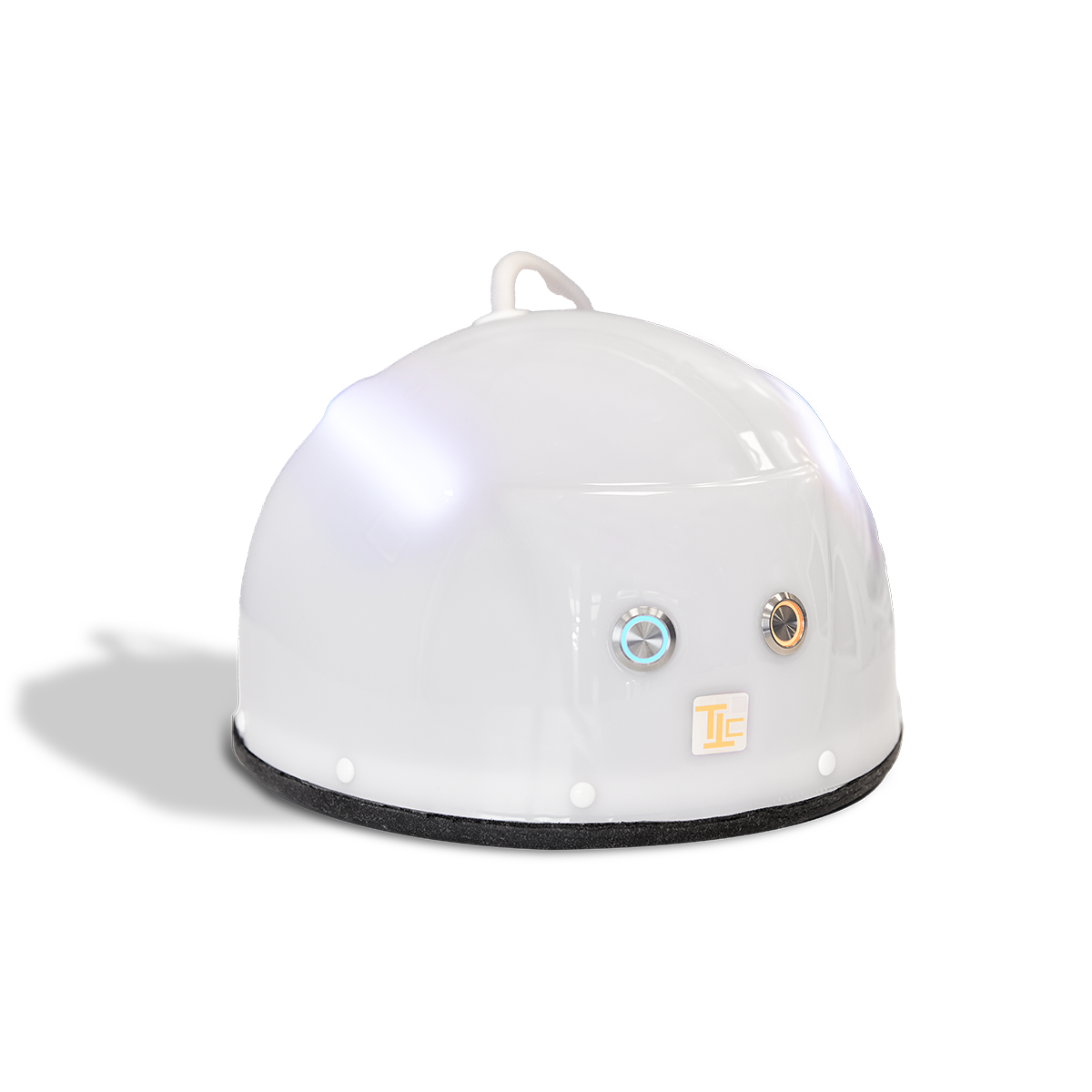
Earlier-generation TES acquisition systems, used in ongoing research and clinical development.

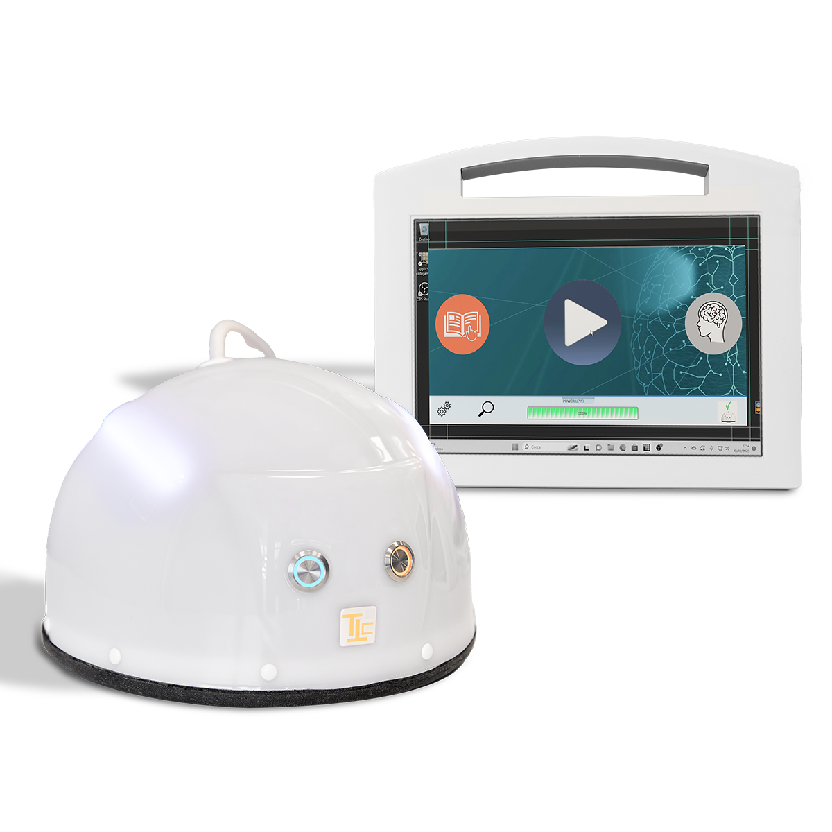
Temnograph TES A90

Temnograph TES A90

Prototype
Temnograph TES A100

Temnograph TES A100

Prototype

TEMNOGRAPHY owns the Temnography® technology, including the related trademarks, patents and know-how. B&B S.r.l. has operated under mandate from TEMNOGRAPHY as manufacturer of the TES HT family and has been responsible for its clinical and regulatory development.